What Are Dental Radiographs and Why Are They Crucial?
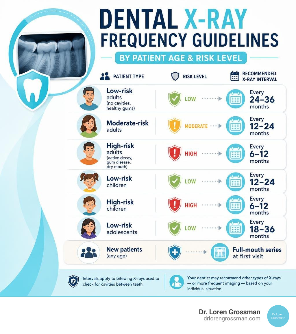
Dental x-rays how often you need them depends on your age, oral health, and risk level — but here are the general guidelines most dentists follow:
| Patient Type | Recommended Interval |
|---|---|
| Low-risk adults (no cavities, healthy gums) | Every 24-36 months |
| Moderate-risk adults | Every 12-24 months |
| High-risk adults (active decay, gum disease, dry mouth) | Every 6-12 months |
| Low-risk children | Every 12-24 months |
| High-risk children | Every 6-12 months |
| Low-risk adolescents | Every 18-36 months |
| New patients (any age) | Full-mouth series at first visit |
These intervals apply specifically to bitewing X-rays used to check for cavities between teeth. Your dentist may recommend other types of X-rays — or more frequent imaging — based on your individual situation.
Dental X-rays are one of the most important tools in dentistry. They reveal problems that a visual exam simply cannot catch — things like decay hiding between teeth, bone loss, infections, and cysts developing silently beneath the gumline. Catching these issues early almost always means simpler, less expensive treatment.
The good news: modern digital X-rays are very safe. A full set of digital X-rays delivers roughly the same radiation as a single day of natural background radiation — less than what you’d absorb on a cross-country flight.
I’m Dr. Loren Grossman, a family and cosmetic dentist serving the Wyoming Valley since 1984, and over four decades of practice I’ve used dental x-rays to detect problems at every stage — from early childhood cavities to complex bone loss in adults — helping patients avoid far more serious procedures down the road. Understanding dental x-rays how often you truly need them is something I discuss with every patient individually, and I’ll walk you through exactly how those decisions are made.
When you sit in the dental chair, our primary focus is keeping your smile healthy, functional, and beautiful. However, during a standard visual examination, we can only see what is happening on the surface of your teeth and gums. To truly understand your oral health, we must look deeper. This is where dental radiographs (X-rays) become indispensable.

Dental X-rays are essential, preventive, and diagnostic tools that pass a highly concentrated beam of electromagnetic energy through the soft tissues of your mouth. Because denser structures like teeth and jawbones absorb more of this energy, they show up clearly as light areas on the image, while cavities, infections, and soft tissues appear darker.
Without this technology, a dentist is essentially working half-blind. Some of the most critical oral health issues develop entirely out of sight, including:
- Hidden Decay: Cavities frequently form on the contact surfaces between teeth where toothbrush bristles and visual tools cannot reach.
- Deep-Rooted Infections and Abscesses: Infections can form at the very tip of a tooth’s root, deep within the jawbone, causing pain or silently destroying bone.
- Bone Loss: Periodontal (gum) disease attacks the bone support holding your teeth in place. X-rays show us the exact height of your jawbone so we can intervene before teeth become loose.
- Cysts and Tumors: Non-cancerous cysts, fluid-filled sacs, and occasionally benign or malignant tumors can develop within the jawbone without causing any early physical symptoms.
- Developmental Abnormalities: For younger patients, X-rays show how adult teeth are forming, whether they are erupting in the correct positions, and if any teeth are missing or impacted.
By utilizing these diagnostic images, we can catch minor issues before they transform into painful, complex, and costly dental emergencies. This proactive approach is the cornerstone of Preventive Dentistry Practices in Pittston: How Regular Visits Can Save Your Smile, ensuring that we preserve your natural tooth structure for as long as possible.
Dental X-Rays How Often: Guidelines for Patients
One of the most common misconceptions is that dental X-rays must be taken on a rigid, calendar-based schedule at every single visit. In modern dentistry, this “one-size-fits-all” approach is outdated. National organizations, including the American Dental Association (ADA) and the Food and Drug Administration (FDA), emphasize that radiographic examinations must be tailored to the individual patient.
Determining dental x-rays how often you need imaging is a clinical decision that we make only after performing a comprehensive clinical examination, reviewing your complete dental and medical history, and conducting a thorough caries risk assessment.
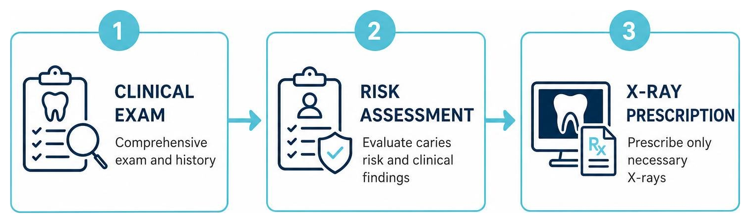
As shown in the clinical guidelines outlined in DENTAL RADIOGRAPHIC EXAMINATIONS: Recommendations for Patient Selection, basing our imaging decisions on active clinical evaluations rather than a strict schedule can lead to up to a 43 percent reduction in radiographs without any clinically consequential increase in undiagnosed dental disease. We only order X-rays when there is a clear diagnostic need, keeping your exposure as low as reasonably achievable.
Dental X-Rays How Often for New vs. Recall Patients
The frequency of your dental X-rays depends heavily on whether you are visiting our practice for the first time or returning for a routine check-up.
New Patients (Establishing a Baseline)
When you join our practice as a new patient, we need to establish a comprehensive baseline of your oral health. This is vital because we need to see what is happening underneath your existing dental work, evaluate your bone levels, and check for any silent, asymptomatic issues.
For a new adult patient, we typically recommend a full-mouth series of radiographs (often called a FMX) or a combination of bitewings and a panoramic X-ray. If you have had recent X-rays taken at another dental office, we always encourage you to have those records transferred to us to avoid duplicating images unnecessarily.
Recall Patients (Routine Monitoring)
Once we have established your baseline and understand your oral health, your recall schedule is customized. Asymptomatic patients with outstanding oral hygiene, no history of cavities, and healthy gum tissue do not need X-rays at every six-month visit. For low-risk adults, bitewing X-rays are perfectly appropriate every 24 to 36 months.
However, if you have a history of frequent cavities, active gum disease, or other ongoing concerns, we will recommend more regular monitoring. Keeping up with these customized intervals is a key highlight of The Importance of Regular Dental Check-Ups in Wilkes-Barre: Insights from Dr. Grossman, where we focus on personalized care schedules.
Risk Factors That Determine Dental X-Rays How Often You Need Imaging
Your risk category is not permanent; it can change over time due to shifts in your lifestyle, diet, age, and medical health. We evaluate several key risk factors to decide if you need more frequent imaging:
- High Caries (Cavity) Risk: If you have had active decay within the last year, have poor dietary habits (such as a high-sugar diet), or struggle with oral hygiene, you may need bitewing X-rays every 6 to 12 months to catch new decay before it reaches the tooth pulp.
- Periodontal Disease: Patients with active gum disease or a history of bone loss require periodic X-rays (usually every 12 to 24 months, or sooner if undergoing active periodontal therapy) to monitor jawbone stability.
- Orthodontic Treatment: If you are wearing braces or clear aligners, we use X-rays to assess tooth movement, monitor root lengths (to ensure the bone movement isn’t causing root resorption), and check the alignment of the jaw.
- Dry Mouth (Xerostomia): A lack of saliva is highly dangerous for teeth. Saliva neutralizes acids and washes away food particles. If you suffer from dry mouth due to medications, Sjögren’s syndrome, or radiation therapy, your risk of rapid cavity development skyrockets, requiring more frequent X-rays.
- Age and Development: As detailed in the clinical guidelines for Prescribing Dental Radiographs for Infants, Children, and Adolescents, children and teenagers have developing mouths where teeth are shifting and jaws are growing rapidly. Because their enamel is thinner, decay can spread to the nerve of a baby tooth much faster than in an adult, often necessitating X-rays every 6 to 12 months if they are prone to cavities.
Types of Dental X-Rays and What They Reveal
Just as a mechanic uses different tools to inspect different parts of an engine, dentists use several distinct types of X-rays to evaluate different aspects of your oral anatomy.

The most common types of imaging we use in our General Dentistry Services include:
- Bitewing X-Rays: These are the standard images taken during routine check-ups. You bite down on a small tab or holder, and the image captures the crowns of your upper and lower teeth in a specific area. These are highly effective for detecting cavities between teeth and monitoring early bone loss around the tooth necks.
- Periapical (PA) X-Rays: Unlike bitewings, which only show the crowns, a periapical X-ray captures the entire tooth from the chewing surface down to the very end of the root and the surrounding jawbone. We use PAs to diagnose toothaches, plan root canals, check for deep abscesses, and monitor dental implants.
- Panoramic X-Rays: This extraoral image is taken by a machine that rotates around your head. It captures your entire mouth in a single, wide 2D image, showing all of your upper and lower teeth, the jaw joints (TMJ), the nasal cavity, and the sinuses. Panoramic X-rays are excellent for evaluating wisdom teeth, identifying jaw fractures, and screening for impacted teeth or deep cysts.
- Cone-Beam Computed Tomography (CBCT): This advanced 3D imaging technology constructs a highly detailed three-dimensional model of your teeth, jawbone, nerve pathways, and sinuses. A CBCT scan is incredibly useful for planning complex treatments like dental implant placements, evaluating cracked teeth that don’t show up on 2D images, and performing precise endodontic (root canal) therapy.
Safety, Radiation Exposure, and Modern Technology
It is completely natural to have questions about radiation safety. However, when we look at the actual scientific data, the amount of radiation exposure from modern dental imaging is exceptionally low.
To put things in perspective, we are exposed to natural background radiation every single day from the soil, rocks, atmosphere, and even cosmic rays. In the United States, the average person receives about 3.1 millisieverts (mSv) of natural background radiation per year.
Let’s compare that to the doses received from modern dental imaging:
- Four digital bitewing X-rays: ~0.002 mSv (equivalent to less than a single day of natural background radiation, or about 1/15th of the exposure from a cross-country flight).
- Full-mouth digital X-ray series: ~0.005 mSv (about 1.5 days of natural background radiation).
- Chest X-ray: ~0.100 mSv (roughly 20 to 50 times more radiation than a set of dental bitewings).
- Annual occupational limit for health care workers: 50 mSv per year.
At our practice, we strictly adhere to the ALARA Principle, which stands for As Low As Reasonably Achievable. This means we take every possible precaution to minimize your exposure:
- Digital Sensors: We use state-of-the-art digital radiography instead of traditional film. Digital imaging provides an outstanding diagnostic image while reducing radiation exposure by 40 to 60 percent compared to older film techniques.
- Rectangular Collimation: The collimator is the device that shapes the X-ray beam. By switching from a traditional circular collimator to a highly targeted rectangular collimator, we restrict the beam precisely to the size of the digital sensor, which decreases the radiation dose by up to fivefold.
- Optimized Exposure Times: We “child-size” our exposure settings for younger patients, reducing the radiation time to match their smaller, less dense bone structures.
- Long Source-to-Skin Distance: Utilizing a longer cone distance (40 cm rather than 20 cm) further reduces unnecessary skin exposure by 10 to 25 percent.
Additionally, our understanding of patient safety continues to evolve. In line with the updated guidelines published in New Recommendations Confirm Dental X-Rays Most Effectively Used in Moderation and the X-Rays/Radiographs | American Dental Association resource, routine patient shielding—such as lead aprons and thyroid collars—is no longer universally recommended for modern digital systems.
Because modern digital beams are so tightly collimated and digital sensors require so little radiation, the amount of scatter radiation reaching the rest of your body is virtually negligible. In fact, large thyroid collars can sometimes accidentally block the primary X-ray beam, requiring us to retake the image and actually increasing your overall exposure. However, we always prioritize your comfort and peace of mind, and we are happy to discuss safety protocols and customize protective steps to make you feel completely secure during your visit.
Frequently Asked Questions About Dental X-Rays
We believe that an informed patient is a healthy, happy patient. Here are some of the most common questions we hear at our Kingston, PA office regarding dental imaging safety and necessity.
Are dental X-rays safe during pregnancy?
Yes, necessary dental X-rays are completely safe during pregnancy. If you are experiencing a toothache, a dental infection, or gum issues while pregnant, obtaining a proper diagnosis is absolutely critical. Untreated dental infections pose a far greater risk to both you and your developing baby than the negligible amount of radiation from a dental X-ray.
Because the X-ray beam is aimed strictly at your head and neck, the amount of scattered radiation reaching your abdomen is practically zero. However, as a matter of standard safety and to protect your peace of mind, we generally defer entirely elective, cosmetic-related X-rays until after your delivery. For more tips on maintaining your oral health during pregnancy, check out Preventive Dental Care Tips from Your Scranton Dentist to Protect Your Oral Health.
Do children need more frequent dental X-rays than adults?
In many cases, yes. Children’s mouths undergo rapid changes as baby teeth are lost and permanent teeth erupt. Additionally, the enamel on primary (baby) teeth is significantly thinner than adult enamel. This means that once a cavity forms between a child’s teeth, it can travel through the enamel and reach the sensitive pulp (nerve) of the tooth much faster than it would in an adult.
To prevent painful tooth infections and premature tooth loss—which can cause severe crowding issues when adult teeth try to erupt—we may recommend X-rays every 6 to 12 months for children who are at a moderate to high risk for tooth decay.
How do dental X-rays save money in the long run?
Think of dental X-rays as an investment in your financial and physical health. A simple set of routine bitewing X-rays can detect a tiny cavity forming between your teeth when it is still small enough to be treated with a conservative, inexpensive dental filling.
If you skip X-rays, that tiny cavity will continue to grow undetected beneath the surface. By the time it finally starts hurting, the decay has likely reached the nerve, requiring a root canal, a protective crown, or even an extraction followed by a costly dental implant. Spending a small amount on preventive imaging today can save you thousands of dollars in extensive restorative treatments down the road.
Conclusion
At the dental practice of Dr. Loren Grossman, we are dedicated to providing the highest standard of personalized cosmetic and general dentistry in Northeastern Pennsylvania. Serving patients in Kingston, Pittston, Wilkes-Barre, Scranton, and the surrounding Wyoming Valley communities, we combine advanced digital imaging technology with a warm, patient-first approach to deliver beautiful, healthy smile transformations.
We never treat X-rays as a routine formality. Every scan we perform is carefully considered, keeping your safety, comfort, and unique oral health needs at the forefront of our care. If you are due for a routine check-up, have questions about your diagnostic needs, or want to discuss a customized treatment plan, we are here to help.
Schedule your comprehensive dental consultation today and let our experienced team help you achieve a lifetime of healthy, confident smiles!